Grounded in peer-reviewed science.
Across three publications since 2024, our team has established that AI can rapidly and accurately identify fungal pathogens from microscope images, demonstrated the approach with additional imaging modalities, and combined explainable AI to “peer inside the black box,” paving the way for regulatory approval.
- 97%
- Top published identification accuracy (C. parapsilosis).
- 3
- Peer-reviewed publications across Medical Mycology, Journal of Imaging, and Scientific Reports.
- 7
- Fungal species identified across the published research panel.
- AICE
- Alberta Innovates Accelerating Innovations in CarE grantee.
Three years. Three peer-reviewed papers.
- 2026Scientific ReportsPeer-reviewed
Guthrie, Shankarnarayan, and Charlebois
Study that demonstrates that advanced computer vision models can identify a wide range of fungal pathogens from microscope images. This research uses explainable AI methods to peer inside the “black box” to reveal the biological/non-biological features machine learning models use to classify pathogens, paving the way for regulatory approval.
Read the paper - 2026Journal of ImagingPeer-reviewed
Liu, Shankarnarayan, Cheng, Gupta, Rozmus, Mandal, Charlebois, and Tsui
Study that demonstrates that CNNs can also be trained to identify the above fungal pathogens with similar speed and accuracy using other modalities (scattered light data).
Read the paper - 2024Medical MycologyPeer-reviewed
Shankarnarayan and Charlebois
Study that establishes that convolutional neural networks (CNNs) can rapidly and accurately identify the species of clinically-relevant fungal pathogens (C. albicans, N. glabratus, C. haemulonii, and the emerging pathogen C. auris) from microscope images.
Read the paper
DOIs and direct links provided on request. Contact info@biosciai.ai.
Peering inside the black box.
For the majority of true-positive predictions, our models place high importance on biologically relevant features. Combining computer vision with explainable AI is what paves the way for regulatory approval.
Highly ranked across true-positive predictions.
A morphology that varies by species.
Internal patterns the model weighs.
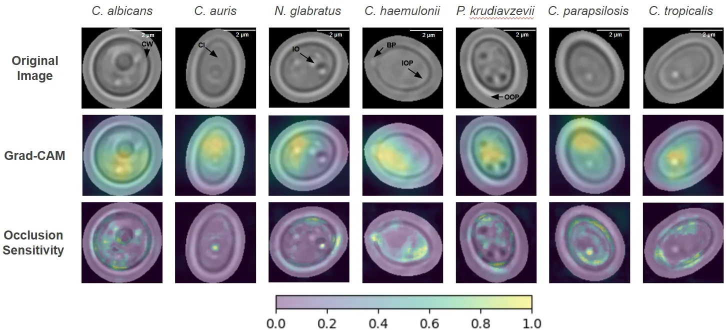
Two independent attribution methods agree on biologically relevant regions across all seven target species.
Signatures specific to each species.
Methods that peer inside the black box.
Transparent decisions for clinical adoption.
Supported by Alberta innovation funding.

Researcher, clinician, or collaborator?
Open to collaboration.