“Why are we still using 20th-century methods for a 21st-century threat?”
Fungal infections, once considered rare, are now a rapidly escalating global health crisis. C. auris has spread to more than 65 countries, with mortality rates reaching up to 60% in vulnerable patients. Yet the diagnostic tools available to hospitals remain slow, expensive, and inaccessible.
We decided to change that.
From clinical sample to species call.
-
Load the sample.
Clinical sample is loaded onto the device.
-
Pathogen isolation.
Proprietary microfluidics technology separates the pathogen from the sample.
-
Image capture.
Microscopy images of the yeast cells are captured.
-
AI analysis & identification.
The trained AI model analyzes the images and returns a species-level identification with a confidence score.
-
Result at bedside.
Results are delivered to the clinician in under an hour, right at the patient's bedside.
From a drop of blood to a named species.
A complete diagnostic workflow built around a portable reader, a single-use cartridge, and machine learning trained on real pathogen data.
Sample in. Identification in under an hour.
A small volume of patient blood is loaded into a single-use microfluidic cartridge that performs automated sample preparation and presents the pathogens for analysis. The portable reader then captures high-resolution optical or sensor data, which is processed in real time by machine learning models to detect and identify the specific organism (including challenging and resistant species such as C. auris), delivering actionable results in under an hour.
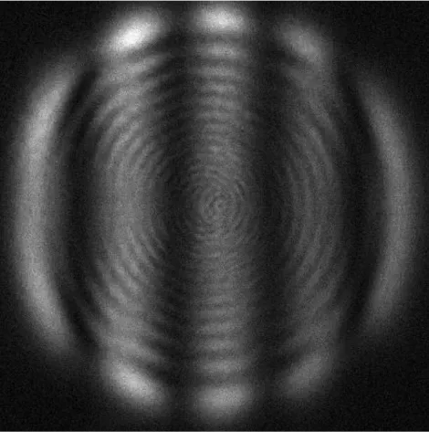
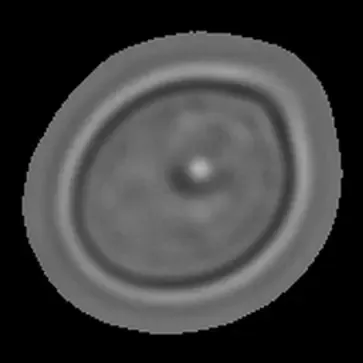
The reader captures raw microscopy and light-scattering images. The AI learns species-specific signatures directly from these inputs.
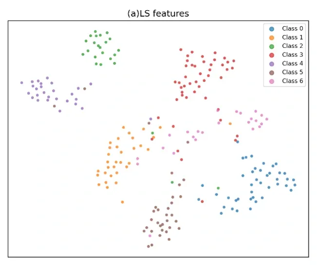
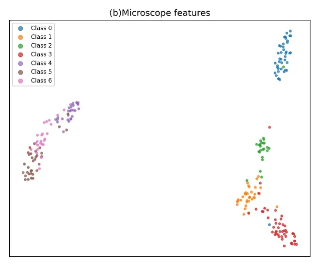
t-SNE projections of learned features. Both modalities produce clean per-species clusters, evidence the model generalizes across data types.
Microfluidics meets deep learning.
The core approach integrates microfluidics for precise, low-volume fluid handling and pathogen enrichment with deep learning algorithms, primarily computer vision models trained on large, curated datasets of pathogen morphology and light-scattering characteristics. Unlike traditional culture-based microbiology or broad molecular assays, the system bypasses lengthy incubation steps by using AI to recognize subtle patterns in the captured data. Continuous model improvement through additional training data further enhances accuracy and expands the range of detectable pathogens over time.
Built for the bedside.
Conventional methods often require 24 to 72 hours and specialized lab infrastructure, delaying targeted antifungal therapy and driving overuse of broad-spectrum drugs. By bringing identification to the point of care, BioSciAI enables earlier, more precise treatment in critical settings such as ICUs and emergency departments, supporting antimicrobial stewardship and better patient outcomes at lower cost.
- Point-of-care speed
- Species-level results in under an hour, at the patient's bedside.
- Species-level resolution
- The exact organism, including emerging and hard-to-culture threats.
- No central lab
- Portable, with minimal operator training required.
Seven species. One platform.
Independent identification-accuracy figures across seven life-threatening fungal pathogens.
Source
Identification accuracy figures provided by BioSciAI from peer-reviewed research. See the research page for full publication details.
- C. parapsilosis WHO High97%
- C. auris WHO Critical90%
- P. kudriavzevii WHO Medium90%
- C. albicans WHO Critical89%
- N. glabratus WHO High85%
- C. tropicalis WHO High82%
- C. haemulonii76%
Faster. Cheaper. At the bedside.
Current diagnostic standards remain slow, expensive, labor-intensive, and heavily dependent on centralized laboratories. These delays directly contribute to postponed antifungal therapy, prolonged hospital stays, and higher mortality.
Bring AI-driven pathogen identification to your institution.
Open to clinical, research, and pharmaceutical partnerships.